
Patient Education Resource

The eye is a very intricate and complex organ containing over 2 million parts. For this reason, there are many sub-specialities in ophthalmology and optometry. Retina specialist are ophthalmologists who specialize in retinal disease. The retina is a very sensitive neuro-tissue that lines the inside of the eye. To make sense of how the eye works, think of the eye as a camera. At the front of the eye is a high-powered lens, and on the inside is the film (the film being the retina). When light enters the eye, the light is focused onto the retina from the lens. In the retina, microscopic cells (cones and rods) are stimulated by light and create chemical signals. These signals travel through the retina until reaching the optic nerve. The optic nerve is a special nerve within the brain that bridges the chemical signals from the retina to the visual cortex. The visual cortex is a specific region in the back of the brain which translates the chemical signals into the images we perceive. The images above is a map that will help you understand the pathway of light.
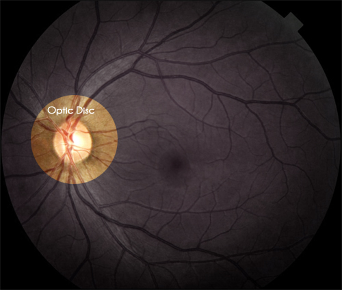
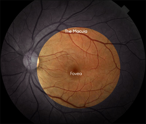
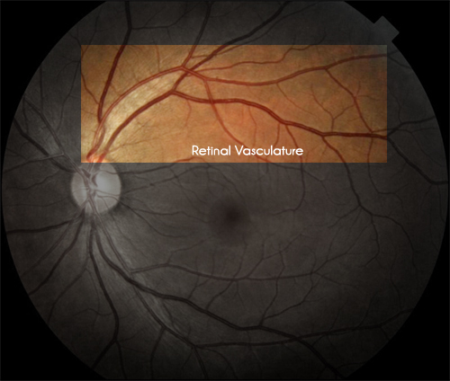
Know Your Retinal Anatomy
Education Categories
Test your understanding of the eye and retina
Additional Questions
Retina
Iris
Elevated eye pressure resulting in damage to the optic nerve
Flashes of light, floaters, darkening, or a curtain/shadow over your vision
While AMD is primarily diagnosed in elderly patient's, the disease can be seen in patient's as young as 55.
Cornea
False. There is a common misconception that cataracts are very dangerous. The truth, however, is that everyone will eventually develop a cataract (just like a wrinkle on your skin). Over time, cataracts can worsen and cause a decline in vision. Once your vision is limited significantly by a cataract, cataract extraction can be performed and a new artificial lens will be placed. As long as your retina is healthy, your vision will be improved significantly following cataract extraction.
Sclera
Fovea
False. There are many treatments for diabetic retinopathy. To ensure you do not develop permanent vision loss, yearly dilated eye exams are strongly advised for any person with diabetes.
Pupil
Uvea/Choroid
Visual Cortex
If your question was not answered here, feel free to call us at (602) 232-6066