
The Retina
The retina is comprised of ten layers, all in which work together to transmit chemical signals to the optic nerve. Ocular Coherence Tomography (OCT) can be performed to evaluate the health of these retinal layers. Obtaining an OCT scan is painless and can be completed in under two minutes. Many retinal conditions can be evaluated through the use of an OCT image, such as: macular degeneration, central serous retinopathy, diabetic macular edema, macular hole, and epiretinal membrane(s).
When come to see us, and we perform an OCT, we will always present your scans to you and explain any abnormalities found and the treatment options available. The image above demonstrates a healthy, normal looking retina on an OCT scan. There are many factors which may change the appearance of your OCT scan in comparison to the image above. For example, your refractive error (myopic changes) can change the curvature of your OCT. Other factors include the machine used in obtaining your OCT scan. In our office, we use machines with the highest resolution available and the latest in technology. Furthermore, the machines used in our office are approved for imaging for clinical trials and research studies.
Retinal Photography
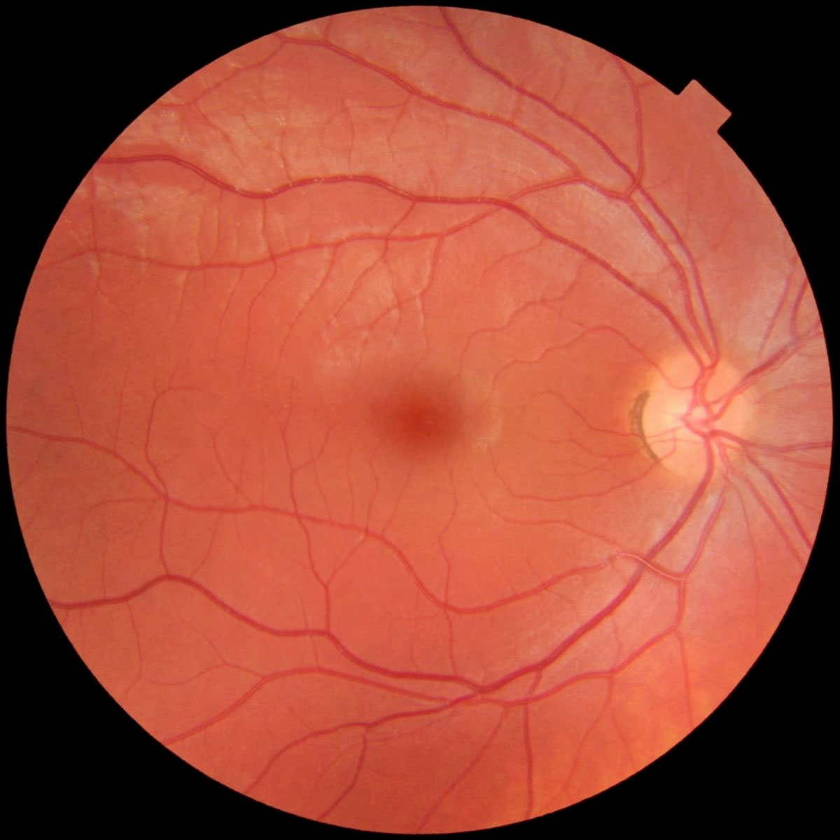
There are many different types of retinal photography including red-free imaging, ICG angiography, fluorescein angiography, and auto-florescence. Each type of image serves a different purpose in evaluating the retina. The image you see to the left is an example of a normal retina being photographed with no filters. This type of photograph allows a large and more detailed inspection of the retina for patient's that have irregular retinal changes. Fundus photography is also often performed in order to keep a baseline of progression or improvement in a patient's retinal condition.
The image to the right is an example of fluorescein angiography (FA). An FA is performed when the ophthalmologist needs to evaluate the blood flow of the retina. When performing an FA, the patient will have a IV placed for approximately 5 minutes at which time fluorescein is injected into a vein on the arm. The fluorescein will travel to the eye at which time the blood vessels within the eye will begin to light up. Fluorescein is non-toxic, however, you should always notify your physician if you are pregnant before performing FA imaging.

Retinal Ultrasonography
An ultrasound of the eye is performed when the ophthalmologist cannot view the retina clearly and needs to rule out a retinal detachment or mass. Additionally, retinal ultrasonography (B-Scan) is performed to monitor progression of ocular tumors or lesions. A B-Scan is painless and can be completed with your eyes closed. A probe will rest on your closed eyelid and an image will be taken. The entire process can take 1 to 2 minutes.
